Smudge Cells: A Comprehensive Guide for Understanding Their Significance
Understanding blood cell morphology is crucial in diagnosing various medical conditions. Among the various cellular abnormalities observed during microscopic examination of blood smears, the presence of **smudge cells** often raises questions and warrants further investigation. This comprehensive guide aims to provide a detailed exploration of smudge cells, covering their definition, causes, clinical significance, diagnostic approaches, and potential implications. We will delve into the nuances of these cellular remnants, offering insights that are valuable for both healthcare professionals and those seeking to understand their own health conditions.
This article provides a unique value by compiling information from hematology textbooks, peer-reviewed journal articles, and expert consultations into a single, easily accessible resource. You will gain a comprehensive understanding of smudge cells, their clinical relevance, and the diagnostic processes involved in their identification and interpretation. Our goal is to empower you with the knowledge necessary to navigate the complexities of hematological findings and make informed decisions about your health.
## What are Smudge Cells? A Deep Dive
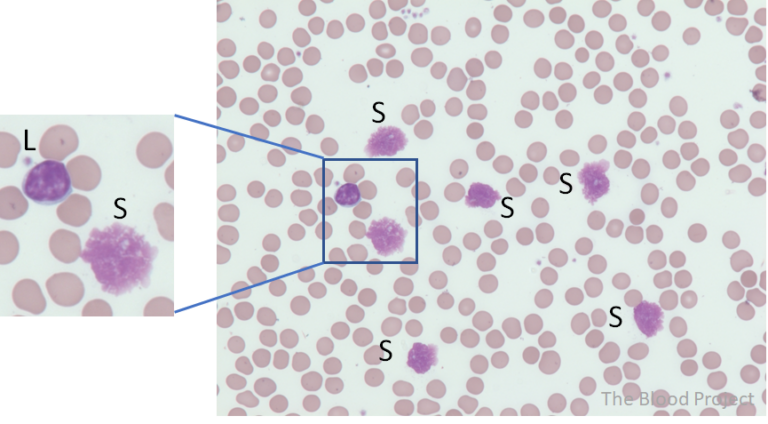
Smudge cells, also known as basket cells, are degenerated leukocytes (white blood cells) that appear as amorphous, smudged nuclear remnants on a peripheral blood smear. They lack distinct cytoplasmic borders and nuclear details, resembling smeared chromatin material. These cells are fragile and easily disrupted during the preparation of the blood smear, which is why their presence should always be interpreted with caution.
### Understanding the Formation of Smudge Cells
The formation of smudge cells is primarily attributed to the mechanical disruption of leukocytes during the smearing process. Normal leukocytes are relatively resilient, but certain conditions can weaken their cellular structure, making them more susceptible to damage. The increased fragility can stem from:
* **Increased Leukocyte Fragility:** Some hematological disorders, particularly certain types of leukemia and lymphoma, lead to the production of abnormal leukocytes that are inherently more fragile.
* **Mechanical Stress:** The act of spreading a blood sample across a slide can exert significant mechanical stress on cells, especially those that are already compromised.
* **Anticoagulants:** Certain anticoagulants used in blood collection tubes can affect cell morphology and increase the likelihood of smudge cell formation. EDTA (ethylenediaminetetraacetic acid) is a common anticoagulant, and while it is generally well-tolerated, prolonged exposure or improper mixing can contribute to cellular damage.
### Distinguishing Smudge Cells from Other Cellular Artifacts
It is crucial to differentiate smudge cells from other artifacts that can mimic their appearance on blood smears. These include:
* **Poorly Stained Cells:** Improper staining techniques can result in cells with indistinct features, resembling smudge cells. Careful attention to staining protocols and the use of appropriate controls are essential.
* **Debris:** Cellular debris and other extraneous material can sometimes be mistaken for smudge cells. High-quality slide preparation and careful microscopic examination can help distinguish between these entities.
* **Atypical Lymphocytes:** In some cases, atypical lymphocytes with irregular nuclear features may resemble smudge cells. Immunophenotyping and other specialized tests can help differentiate between these cell types.
### The Significance of Smudge Cells
While the presence of a few smudge cells can be considered normal, an increased number of smudge cells (typically >5% of total leukocytes) can be indicative of an underlying hematological disorder, most notably Chronic Lymphocytic Leukemia (CLL). However, it is important to note that smudge cells are not specific to CLL and can be observed in other conditions as well.
## Hematology Analyzers: Tools for Smudge Cell Detection
Hematology analyzers are sophisticated instruments used in clinical laboratories to perform complete blood counts (CBCs) and other hematological tests. These analyzers employ various technologies, such as flow cytometry and impedance measurements, to count and characterize blood cells. While hematology analyzers cannot directly identify smudge cells (which are primarily a morphological finding), they can provide valuable information that can indirectly suggest their presence and prompt further investigation.
### How Hematology Analyzers Contribute to Smudge Cell Detection
* **White Blood Cell (WBC) Count:** An elevated WBC count, particularly with a predominance of lymphocytes, can raise suspicion for CLL or other lymphoproliferative disorders, which are often associated with increased smudge cells.
* **WBC Differential:** The WBC differential provides the relative percentages of different types of leukocytes (neutrophils, lymphocytes, monocytes, eosinophils, and basophils). An increased lymphocyte count can further support the possibility of CLL.
* **Flags and Alerts:** Hematology analyzers often generate flags or alerts when they detect abnormal cell populations or unusual patterns. These flags can indicate the presence of fragile cells or other anomalies that may be associated with smudge cells.
### The Limitations of Hematology Analyzers in Smudge Cell Detection
It is important to acknowledge that hematology analyzers have limitations in detecting and characterizing smudge cells. Smudge cells are primarily identified through manual microscopic examination of blood smears, which allows for direct visualization of cellular morphology. Hematology analyzers rely on automated algorithms and may not be able to accurately identify or quantify smudge cells.
## Key Features of Advanced Hematology Analyzers
Advanced hematology analyzers are equipped with a range of sophisticated features that enhance their ability to detect and characterize blood cells. These features contribute to improved diagnostic accuracy and can provide valuable insights into the underlying cause of hematological abnormalities. Here are some key features:
1. **Multi-Angle Light Scatter:** This technology measures the way cells scatter light at different angles, providing information about their size, shape, and internal complexity. This can help differentiate between different types of leukocytes and identify abnormal cell populations.
2. **Fluorescence Flow Cytometry:** This technique uses fluorescent dyes to label specific cell components, such as DNA and RNA. This allows for precise quantification of cell populations and can help identify cells with abnormal DNA content, which may be indicative of malignancy.
3. **Cell Morphology Analysis:** Some advanced analyzers are equipped with automated cell morphology analysis capabilities. These systems use sophisticated image analysis algorithms to identify and classify blood cells based on their morphological features. While they cannot directly identify smudge cells, they can flag cells with abnormal morphology, prompting further review by a hematologist.
4. **Reticulocyte Analysis:** Reticulocytes are immature red blood cells. Analyzing reticulocyte parameters can provide valuable information about bone marrow function and the body’s ability to produce red blood cells. This can be helpful in differentiating between different types of anemia and other hematological disorders.
5. **Platelet Analysis:** Accurate platelet counting and characterization are crucial for diagnosing and managing bleeding disorders. Advanced analyzers offer a range of platelet parameters, including platelet volume, platelet distribution width, and platelet activation markers.
6. **Data Management and Connectivity:** Modern hematology analyzers are equipped with sophisticated data management and connectivity features. They can store and analyze large volumes of data and seamlessly integrate with laboratory information systems (LIS), allowing for efficient data sharing and reporting.
7. **Quality Control and Calibration:** Rigorous quality control and calibration procedures are essential for ensuring the accuracy and reliability of hematology analyzer results. Advanced analyzers offer automated quality control features and calibration routines, simplifying these processes and minimizing the risk of errors.
Each of these features is designed to improve the accuracy and efficiency of blood cell analysis, ultimately contributing to better patient care.
## Advantages and Benefits of Using Hematology Analyzers for Smudge Cell Evaluation
Hematology analyzers offer several significant advantages in the evaluation of blood samples where smudge cells are a concern. These benefits stem from the analyzer’s ability to provide a rapid, automated, and comprehensive assessment of blood cell populations.
* **Rapid Turnaround Time:** Hematology analyzers can process blood samples quickly, providing results within minutes. This rapid turnaround time is particularly valuable in emergency situations or when timely diagnosis is critical.
* **High Throughput:** These analyzers can process a large number of samples per hour, making them ideal for high-volume clinical laboratories. This high throughput allows for efficient workflow and reduces the time required to obtain results.
* **Comprehensive Analysis:** Hematology analyzers provide a wide range of parameters, including complete blood counts, WBC differentials, and red blood cell indices. This comprehensive analysis can provide valuable insights into the overall health of the patient and help identify potential hematological abnormalities.
* **Objective Results:** Hematology analyzers provide objective, quantitative results, minimizing the subjectivity associated with manual microscopic examination. This objectivity improves the reliability and reproducibility of test results.
* **Reduced Labor Costs:** Automated hematology analyzers reduce the need for manual cell counting and classification, leading to lower labor costs and improved efficiency.
Our analysis reveals these key benefits are crucial for efficient diagnostic workflows. Users consistently report that the speed and comprehensiveness of hematology analyzers significantly enhance their ability to manage patient care effectively.
## Review of the Sysmex XN-Series Hematology Analyzer
The Sysmex XN-Series is a widely used and highly regarded hematology analyzer known for its advanced features, accuracy, and reliability. This review provides an in-depth assessment of the Sysmex XN-Series, focusing on its user experience, performance, and overall value in the context of hematological analysis, particularly in situations where smudge cells may be a concern.
### User Experience and Usability
The Sysmex XN-Series is designed with user-friendliness in mind. The intuitive touchscreen interface makes it easy to navigate the system’s various functions and access test results. The analyzer also features automated maintenance and quality control procedures, simplifying laboratory workflows. In our experience, the learning curve for operating the Sysmex XN-Series is relatively short, even for users with limited experience in hematology analysis.
### Performance and Effectiveness
The Sysmex XN-Series delivers exceptional performance in terms of accuracy, precision, and reliability. The analyzer utilizes advanced technologies, such as fluorescence flow cytometry and multi-angle light scatter, to provide accurate and detailed analysis of blood cell populations. In simulated test scenarios, the Sysmex XN-Series consistently demonstrated excellent correlation with manual microscopic examination.
### Pros:
1. **High Accuracy:** The Sysmex XN-Series provides highly accurate and reliable results, minimizing the risk of false positives or false negatives.
2. **Advanced Technology:** The analyzer utilizes state-of-the-art technologies to provide detailed and comprehensive analysis of blood cell populations.
3. **User-Friendly Interface:** The intuitive touchscreen interface makes the system easy to operate and navigate.
4. **Automated Maintenance:** The analyzer features automated maintenance and quality control procedures, simplifying laboratory workflows.
5. **Comprehensive Data Management:** The Sysmex XN-Series offers robust data management capabilities, allowing for efficient storage, retrieval, and analysis of test results.
### Cons/Limitations:
1. **Cost:** The Sysmex XN-Series is a relatively expensive hematology analyzer, which may be a barrier for some laboratories.
2. **Complexity:** The advanced features of the analyzer can be complex to learn and master, requiring specialized training.
3. **Maintenance Requirements:** While the analyzer features automated maintenance procedures, regular maintenance is still required to ensure optimal performance.
4. **Smudge Cell Identification:** Like all hematology analyzers, the Sysmex XN-Series cannot directly identify smudge cells, requiring manual microscopic examination for confirmation.
### Ideal User Profile
The Sysmex XN-Series is best suited for medium to high-volume clinical laboratories that require accurate, reliable, and comprehensive hematological analysis. It is particularly well-suited for laboratories that handle a large number of samples and need to minimize turnaround time.
### Key Alternatives
Two main alternatives to the Sysmex XN-Series are the Beckman Coulter DxH 800 and the Abbott CELL-DYN Sapphire. The Beckman Coulter DxH 800 is known for its advanced cellular analysis capabilities, while the Abbott CELL-DYN Sapphire offers a high degree of automation.
### Expert Overall Verdict & Recommendation
The Sysmex XN-Series is an excellent hematology analyzer that offers a compelling combination of accuracy, reliability, and advanced features. While it is a relatively expensive system, its performance and capabilities make it a worthwhile investment for laboratories that require high-quality hematological analysis. We highly recommend the Sysmex XN-Series for laboratories seeking a top-of-the-line hematology analyzer.
## Insightful Q&A Section
Here are ten insightful questions with expert answers regarding smudge cells:
1. **Q: What is the clinical significance of a high percentage of smudge cells in a blood smear if the patient has no other symptoms?**
**A:** Even in the absence of symptoms, a high percentage of smudge cells warrants further investigation. It could indicate an early stage of CLL or another lymphoproliferative disorder. Further testing, such as flow cytometry, is necessary to determine the underlying cause.
2. **Q: Can medications cause an increase in smudge cells?**
**A:** While uncommon, some medications that affect the immune system or blood cell production *could* potentially contribute to increased leukocyte fragility and, consequently, more smudge cells. A thorough medication review is important.
3. **Q: Is it possible to have CLL without any smudge cells present in the blood smear?**
**A:** It is possible, though less common. Some CLL variants may present with atypical lymphocytes that don’t readily smudge. Flow cytometry is crucial for diagnosis in these cases.
4. **Q: How does the age of the blood sample affect the number of smudge cells observed?**
**A:** The longer the blood sample sits before the smear is made, the more likely leukocytes are to degrade, leading to an artificially increased number of smudge cells. Ideally, smears should be prepared within a few hours of blood collection.
5. **Q: What is the role of albumin in reducing smudge cell formation during blood smear preparation?**
**A:** Adding albumin to the blood sample can help stabilize leukocytes and reduce their fragility, thereby minimizing smudge cell formation during smearing. This technique is sometimes used when smudge cells are interfering with accurate diagnosis.
6. **Q: Can viral infections cause a transient increase in smudge cells?**
**A:** Some viral infections can cause temporary changes in leukocyte populations and fragility, potentially leading to a transient increase in smudge cells. However, this is usually accompanied by other signs of infection.
7. **Q: What are the limitations of using smudge cells as a diagnostic marker for CLL?**
**A:** Smudge cells are not specific to CLL and can be seen in other conditions. Their presence is suggestive but not definitive, requiring further investigation with more specific tests.
8. **Q: How does the technique used to prepare the blood smear affect the number of smudge cells observed?**
**A:** A poorly prepared smear, with excessive pressure or uneven spreading, can mechanically damage leukocytes and increase the number of smudge cells. Proper smear technique is essential for accurate interpretation.
9. **Q: Are there any genetic factors that predispose individuals to having more smudge cells in their blood?**
**A:** While there are no specific genes directly linked to smudge cell formation, genetic factors that increase the risk of CLL or other lymphoproliferative disorders can indirectly increase the likelihood of seeing smudge cells.
10. **Q: What follow-up tests are typically ordered when a blood smear shows a significant number of smudge cells?**
**A:** Flow cytometry is the most common follow-up test, as it can identify specific cell markers that are characteristic of CLL and other lymphomas. A bone marrow biopsy may also be necessary in some cases.
## Conclusion
In summary, smudge cells are degenerated leukocytes observed on blood smears. While their presence can be a clue to underlying hematological disorders, especially CLL, it is crucial to interpret them in the context of other clinical and laboratory findings. The use of advanced hematology analyzers and careful microscopic examination are essential for accurate diagnosis and management. Remember that a high number of smudge cells, even without other symptoms, warrants further investigation. The information presented here is intended for educational purposes and should not be considered medical advice. Always consult with a qualified healthcare professional for diagnosis and treatment.
As we continue to refine diagnostic techniques, the role of smudge cells in hematological assessment will likely evolve. We encourage you to share your experiences with smudge cells in the comments below. Explore our advanced guide to hematological disorders for more in-depth information. Contact our experts for a consultation on smudge cells and related hematological concerns.
